Comparison of MRI slices at the mid calf showing the anatomy with an IP
By A Mystery Man Writer
Description
![Comparison of MRI slices at the mid calf showing the anatomy with an IP](https://media.springernature.com/lw685/springer-static/image/chp%3A10.1007%2F978-3-031-39959-6_5/MediaObjects/606800_1_En_5_Fig20_HTML.jpg)
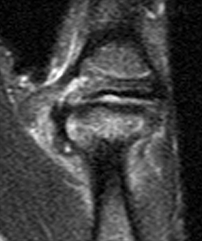
Patella SpringerLink
![Comparison of MRI slices at the mid calf showing the anatomy with an IP](https://static.cambridge.org/binary/version/id/urn:cambridge.org:id:binary-alt:20211124121727-49932-mediumThumb-44331fig5_34c.jpg?pub-status=live)
Applications of radiological imaging in endocrine disease (5C) - Endocrine Pathology
![Comparison of MRI slices at the mid calf showing the anatomy with an IP](https://www.researchgate.net/publication/259701037/figure/fig5/AS:392599820095499@1470614499908/Volume-reduction-of-the-veins-with-different-pressures-exerted-by-a-compression.png)
Volume reduction (%) of the veins with different pressures exerted by a
![Comparison of MRI slices at the mid calf showing the anatomy with an IP](https://www.researchgate.net/profile/Michel-Perrin-3/publication/269639968/figure/fig1/AS:360421342105601@1462942552601/Post-thrombotic-l-femoral-vein-with-fibrotic-lesion-and-thickening-of-the-vein-wall_Q320.jpg)
Comparison between right and left limbs.
![Comparison of MRI slices at the mid calf showing the anatomy with an IP](https://radsource.us/wp-content/uploads/2016/01/1a.jpg)
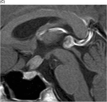
Ulnar Collateral Ligament Tears of the Thumb
![Comparison of MRI slices at the mid calf showing the anatomy with an IP](https://www.researchgate.net/profile/Aikeremujiang-Muheremu/publication/361342437/figure/fig2/AS:1167651334299649@1655401180125/The-hemodynamic-values-are-detected-by-CDFI_Q320.jpg)
Comparative graph depicting the decline in MASI score in both groups
![Comparison of MRI slices at the mid calf showing the anatomy with an IP](https://www.researchgate.net/publication/360777953/figure/fig2/AS:11431281176528516@1690197800794/The-images-are-from-a-three-dimensional-episcopic-dataset-prepared-from-a-human-embryo-at_Q320.jpg)
Comparison of MRI slices at the mid calf showing the anatomy with an IP
![Comparison of MRI slices at the mid calf showing the anatomy with an IP](https://prod-images-static.radiopaedia.org/images/17369613/9da2f7721621e4eeede955d158dfdb_big_gallery.jpeg)
Thoracic spine protocol (MRI), Radiology Reference Article
![Comparison of MRI slices at the mid calf showing the anatomy with an IP](https://www.mdpi.com/applsci/applsci-13-07303/article_deploy/html/images/applsci-13-07303-g001.png)
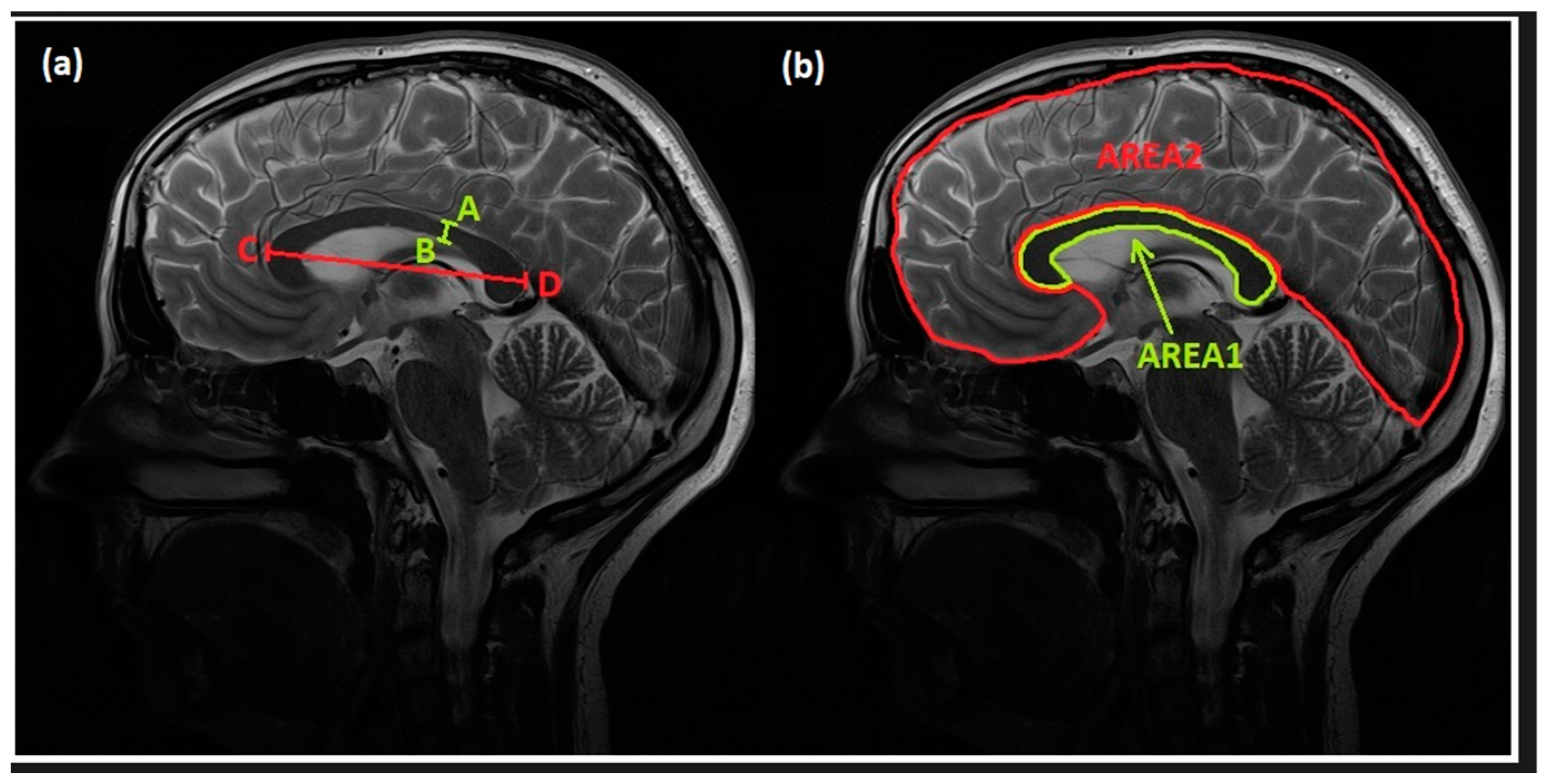
Applied Sciences, Free Full-Text
![Comparison of MRI slices at the mid calf showing the anatomy with an IP](https://www.researchgate.net/publication/23268943/figure/tbl1/AS:667221701427203@1536089458153/58_Q320.jpg)
The occurrence and prevalence of pathological reflux in segments
![Comparison of MRI slices at the mid calf showing the anatomy with an IP](https://www.frontiersin.org/files/Articles/1009411/fcvm-10-1009411-HTML/image_m/fcvm-10-1009411-g001.jpg)
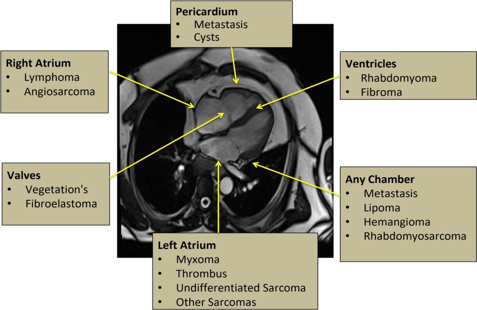
Frontiers Tissue characterization of benign cardiac tumors by cardiac magnetic resonance imaging, a review of core imaging protocol and benign cardiac tumors
![Comparison of MRI slices at the mid calf showing the anatomy with an IP](https://www.researchgate.net/profile/Bert-Ramshorst/publication/14309468/figure/tbl3/AS:704796549988356@1545048000745/The-occurrence-and-prevalence-of-pathological-reflux-in-segments-prevoiusly-affected-by_Q320.jpg)
The occurrence and prevalence of pathological reflux in segments
from
per adult (price varies by group size)