Umbilical hernia, Radiology Case
By A Mystery Man Writer
Description
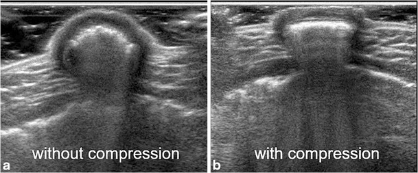
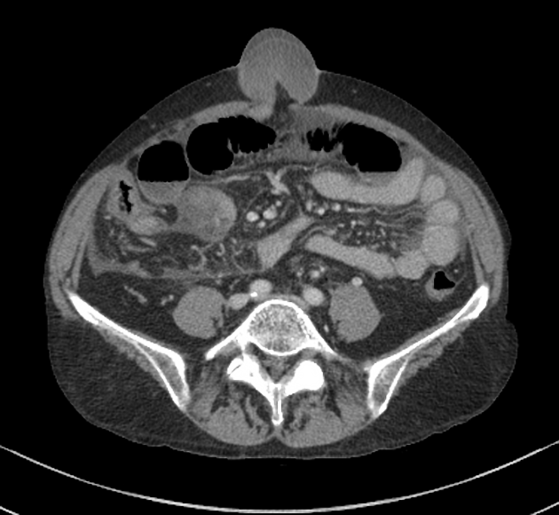
A middle-aged obese male presented with painful umbilical swelling for the last few hours. The ultrasound shows an irreducible umbilical hernia with reduced flow in the herniated small bowel along

Non-contrast 'crunching'-phase CT. These images show a case of
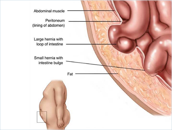
Umbilical hernia presenting as an everted umbilicus in a 7-month-old
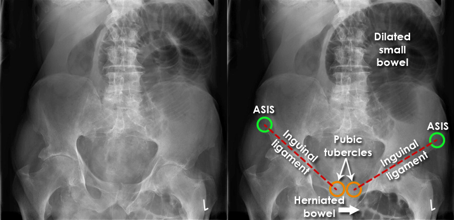
Abdominal X-ray - Abnormalities - Hernias

Abdominal Wall Hernias: Imaging Features, Complications, and Diagnostic Pitfalls at Multi–Detector Row CT
Standardizing the Approach to Hernia Radiology
The Radiology Assistant : Abdominal wall hernias
Five Rare Abdominal Wall Hernias in One Patient: A Case Report - Clinical Case Reports Journal (ISSN 2767-0007)
EPOS™

Abdominal Wall Hernias: Imaging Features, Complications, and Diagnostic Pitfalls at Multi–Detector Row CT

Huge umbilical hernia ultrasound video
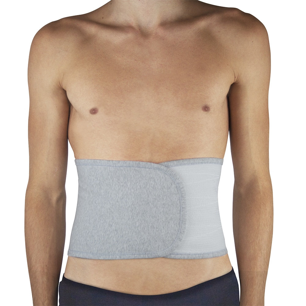
Umbilical hernia, Radiology Reference Article

Hernia Ultrasound Reporting Direct/Indirect Inguinal/Epigastric/Femoral Hernia USG Reports
from
per adult (price varies by group size)